
Artificial Intelligence
Artificial intelligence for radiology in your DICOM viewer!
Medical specialists can have access to a suite of artificial intelligence tools integrated directly into their work environment, allowing them to optimize patient care through predictive and personalized medicine.
Artificial intelligence helps automate specialized tasks and allows radiologists to focus on higher priority diagnostic situations.
- Fully integrated solution
- Certified AI
- Fast image analysis
- Intuitive user experience
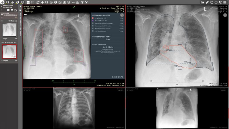
Figure Dicompass - AI description display - Rayscape CXR
We offer simple and fast testing of Artificial Intelligence for automatic description or classification of radiological examinations in your specialized clinics and medical facilities.
We have fully integrated artificial intelligence from these leaders for all our customers, from large regional solutions to small ambulances through these products:
- Dicompass Cloud - smaller clinics and private ambulances
- Dicompass Gateway - hospitals and large healthcare facilities
Integrated Artificial Intelligence:
Carebot
Carebot is an advanced AI tool developed in collaboration with dozens of radiologists to ensure ease of use. It offers exceptional accuracy in detecting even small pathologies, with all data securely anonymized within the healthcare facility. It is available in multiple languages, making it accessible to healthcare professionals worldwide.
Carebot analyzes chest X-rays using artificial intelligence. This unique software accurately detects the following findings: pulmonary lesion, consolidation, pneumothorax, pleural effusion, enlarged cardiac shadow, atelectasis and subcutaneous emphysema.
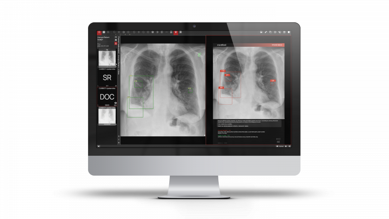
- For each X-ray image, a text evaluation is automatically generated, which can be used as the basis for a medical report.
- Carebot's artificial intelligence can identify all basic radiological findings, including heart shadow measurements. Compared to the competition, it can also detect small, hard-to-detect pathologies.
- Carebot allows diagnostic findings to be displayed as an overlay on the original image in full quality. This overlay can be easily turned on or off with a single click.
- All images analyzed by Carebot are always anonymized on the hospital side, ensuring that patient data never leaves the secure, closed network of the healthcare facility.
- Carebot allows for the filtering of high-risk examinations directly in the PACS environment, enabling the prioritization of patients with potential findings.
More information on the manufacturer's website: carebot.com
Breast-SlimView®
This artificial intelligence is used as clinical decision support system for 2D and 3D mammography. Breast-SlimView® offers an innovative and disruptive reading support where only relevant information is displayed. Radiologists will be able to free themselves from overloaded information and visualize potentially suspicious areas at a glance.
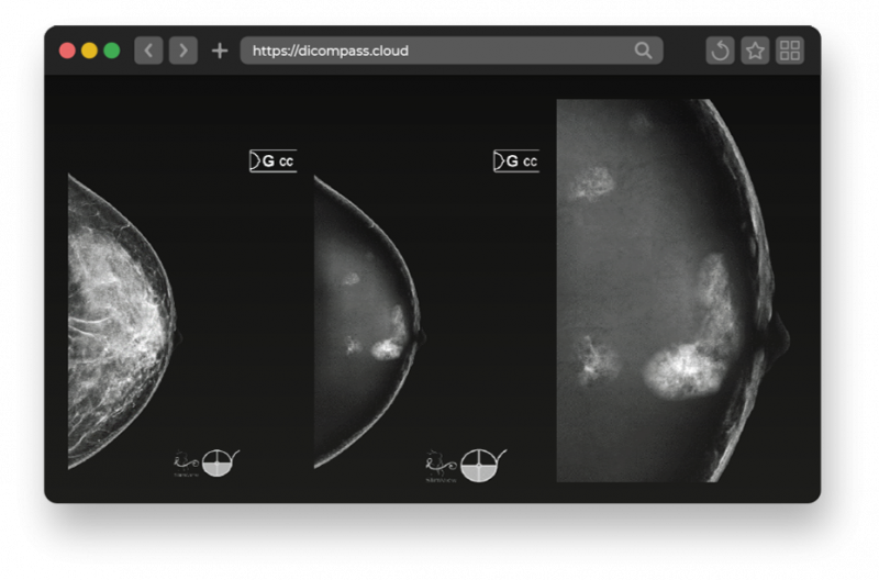
The following software tools are available within this AI to support diagnostics:
- Replacing the uninterested areas with synthetic tissue and keeping only the suspected areas
- Optional evaluation of breast density using the BI-RADS scale, also called Breast Density Evaluation
- Creation of sub-synthetic images for breast tomosynthesis
More information on the manufacturer's website: hera-mi.com
SmartUrgences®
SmartUgences® artificial intelligence is trained to detect abnormalities in all anatomical regions except those listed as contraindications. It focuses on the appendicular skeleton, thorax and pelvis. Its primary purpose is for everyday use in the emergency patient care.
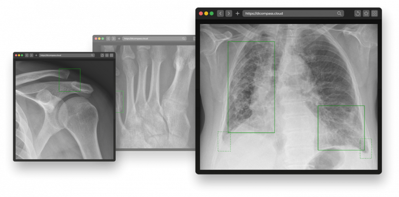
It includes the following pathologies, osteoarticular and pulmonary:
| Fractures | Dislocations | |
| Joint effusions | Pneumothorax | |
| Pulmonary Opacities | Pleural Effusions | |
| Pulmonary Nodules |
More information on the manufacturer's website: milvue.com/solutions/smart-urgences
SmartXpert®
It is designed for efficient and reliable preparation of measurements in musculoskeletal X-ray images. With full integration into our product, these measurements can be further processed. Thus, the artificial intelligence prepares the measurements and the physician can quickly and easily adjust these measurements if necessary.
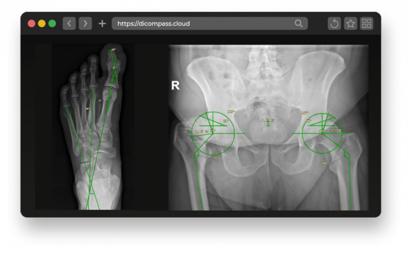
| Gonometry | Hallux varus and hallux valgus | Critical shoulder angle (CSA) | |
| Coxometry | Angles and localisations | Cobb angle | |
| Pelvic tilt | Djian-Annonier angle | Classification of scoliosis | |
| Dysplasia of the hip joint |
qER
This artificial intelligence is designed for find abnormalities on a non-contrast head CT scan, making it a useful tool for diagnosing and evaluating various brain conditions and providing physicians with valuable information for faster diagnosis.
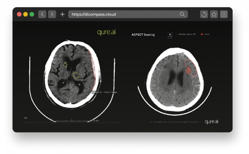
List of abnormalities:
- Intracranial Hemorrhages: Epidural, Subdural, Subarachnoid, Intraparenchymal and Intraventricular hemorrhage
- Cranial Fractures
- Mass Effect: A visible compression or displacement of adjacent structures
- Midline Shift
More information on the manufacturer's website: qure.ai/product/qer
Rayscape CXR
Thanks to artificial intelligence, Rayscape can recognize 148 abnormalities in chest X-rays and group them into 17 different pathological categories. It improves patient care by providing physicians with a quick way to classify and prioritize each patient case.
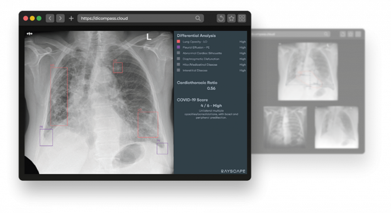
- 148 abnormalities in 17 different pathological classes
- locates groups of pathologies on an X-ray
- generates additional images with subtraction and suppression of bone tissue
- automatically calculates the cardiothoracic index
- identifies SARS-CoV-2 related pathologies and assigns a specific score to each of them
- prioritises patients according to identified pathologies
List of categories:
| Lung Opacity | Atelectasis | Abnormal cardiac silhouette | Consolidation |
| Edema | Diaphragmatic Disfunction | Emphysema | Fracture |
| Hilar/Mediastinal Disease | Interstitial Disease | Lung Lesion | Pleural Effusion |
| Pleural Other | Pneumothorax | Cifo-scoliosis | Support Devices |
| Tuberculosis |
More information on the manufacturer's website: rayscape.ai/chest-xray
Rayscape Lung CT
The Rayscape system helps to identify risk patients and alerts physicians to the presence of possible pathological lesions during regular check-ups. It also enables seamless process optimization, from preventive monitoring and early detection, to emergency care, to tracking the development of changes over time and response to treatment.
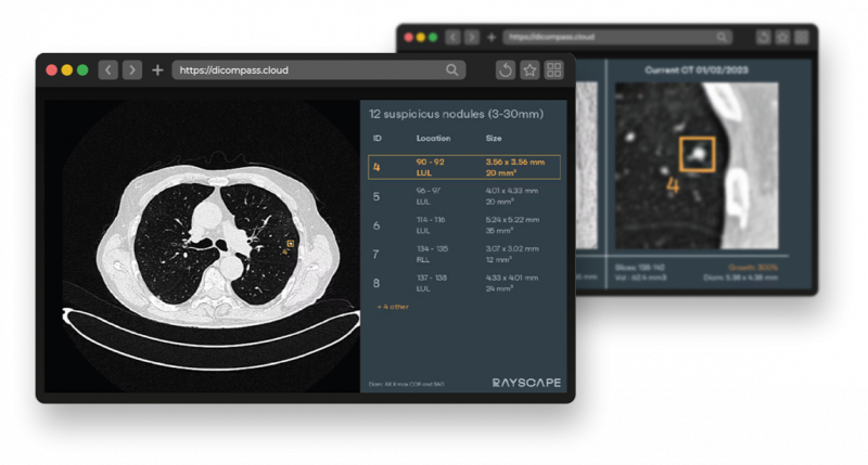
- identifies pulmonary nodules 3-30 mm in diameter
- highlights the presence of nodules on each section and shows their section and lobar location
- automatically measures the diameter and volume of identified nodules
- compares the characteristics of pulmonary nodules between different examinations and generates a final report with nodule progression
- classifies nodules as malignant/benign and assigns a set of attributes including texture, calcification, margins and location to each nodule
- identifies lesions caused by SARS-CoV-2 infection
- displays the degree of lung damage following SARS-CoV-2 infection
More information on the manufacturer's website: rayscape.ai/lung-ct